Overview of Anatomy and Physiology Lab Practical 1
Anatomy and Physiology Lab Practical 1 introduces foundational concepts, including anatomical terminology, cell structure, and histology. Students assess their understanding of key biological structures and processes through hands-on identification and theoretical questions, ensuring a strong foundation for advanced study.
Lab Practical 1 introduces students to foundational anatomical and physiological concepts, focusing on key structures and processes. It assesses understanding of terminology, histology, and microscopy, preparing students for hands-on identification and theoretical application. This practical exam emphasizes critical thinking and precise knowledge, ensuring a solid grasp of essential biological principles.
1.2 Key Objectives and Expectations
Lab Practical 1 aims to assess students’ ability to identify and describe key anatomical structures, understand histological preparations, and demonstrate proficiency in microscopy. Students are expected to apply anatomical terminology accurately, think critically, and manage time effectively during the exam. Mastery of foundational concepts and practical skills is essential for success in this comprehensive assessment.
Anatomical Terminology
Anatomical terminology forms the language of anatomy, comprising directional terms, body planes, and positional descriptions. It provides a standardized way to describe body structures and their relationships, ensuring clear communication in scientific and clinical contexts.
2.1 Key Terms and Definitions
Anatomical terminology includes essential terms like superior, inferior, anterior, posterior, medial, lateral, proximal, distal, dorsal, ventral, and transverse. These terms define relative positions and directions within the body, enabling precise communication about structures and their relationships. Understanding these definitions is critical for accurately identifying and describing anatomical landmarks, ensuring clarity in both theoretical and practical applications of anatomy and physiology studies.
2.2 Directional Terms and Body Planes
Directional terms such as superior, inferior, anterior, posterior, medial, lateral, proximal, distal, dorsal, ventral, and transverse are vital for locating body structures. Body planes, including sagittal, frontal (coronal), and transverse (horizontal), divide the body into sections, aiding in precise anatomical descriptions. Mastery of these terms and planes is essential for accurate identification and communication in anatomy and physiology lab practicals.
2.3 Understanding Anatomical Positions
The standard anatomical position involves standing upright with arms at the sides and palms forward. Directional terms like superior and inferior describe location relative to this position. Body planes, such as sagittal, frontal, and transverse, divide the body into sections, aiding in precise descriptions. Understanding these positions and planes is critical for accurately identifying and locating structures during lab practicals and clinical applications.
Histology and Microscopy
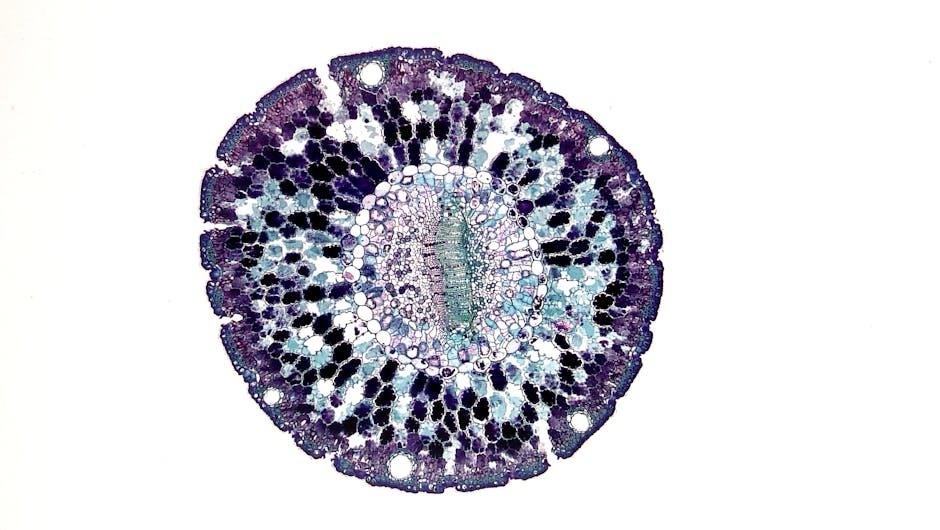
Histology involves studying tissue structure under a microscope, identifying cells, and understanding their organization. Microscopy skills are essential for analyzing slides, recognizing tissue types, and correlating structure with function.
Histology is the study of tissue structure, emphasizing how cells organize into functional units. It involves examining tissue samples under a microscope to identify cell types and arrangements. This field is crucial for understanding tissue function, development, and pathology. In lab practicals, students learn to distinguish between epithelial, connective, muscle, and nervous tissues, correlating their structure with physiological roles.
3.2 Types of Epithelial Tissues
Epithelial tissues are classified into simple and stratified types, based on layer arrangement. Simple epithelium, like squamous, cuboidal, and columnar, consists of a single layer, while stratified epithelium, such as the squamous type, has multiple layers. Each type specializes in functions like diffusion, protection, or secretion, with specific locations and adaptations throughout the body, essential for maintaining barrier and absorptive functions.
3.3 Connective Tissue and Its Functions
Connective tissue supports, binds, and protects body structures. It includes loose, dense, cartilage, bone, and adipose tissue. Functions range from providing structural frameworks to storing energy reserves. Blood, a liquid connective tissue, transports nutrients and oxygen. Each type specializes in maintaining tissue integrity, facilitating movement, and enabling bodily functions, showcasing its vital role in overall physiological processes and organ systems;
Surface Anatomy and Body Regions
Surface anatomy involves identifying external landmarks, such as the sternum, xiphoid process, and costal margins. It helps in understanding body regions and their clinical significance.
4.1 Identifying Surface Landmarks
Identifying surface landmarks involves locating palpable structures like the sternum, xiphoid process, and costal margins. These landmarks guide procedures such as chest compressions or thoracocentesis. Students learn to map external features to internal anatomy, enhancing clinical correlation and diagnostic skills. Proper identification ensures accurate physiological assessments and informed decision-making in medical practices.
4.2 Divisions of the Abdominal and Thoracic Cavities
The abdominal cavity is divided into nine regions: epigastric, umbilical, hypogastric, and four flank regions. The thoracic cavity contains the pleural and pericardial spaces. Understanding these divisions aids in diagnosing conditions like appendicitis or pneumonia. Accurate identification of regions is crucial for clinical assessments and surgical interventions.
The Cell
The cell is the basic structural and functional unit of life, consisting of a plasma membrane, cytoplasm, and organelles. It performs essential functions like metabolism and reproduction.
5.1 Cell Structure and Organelles
The cell consists of a plasma membrane enclosing cytoplasm, where organelles like mitochondria, ribosomes, endoplasmic reticulum, and the Golgi apparatus perform specialized functions. The nucleus houses genetic material, regulating cellular activities. Understanding these structures is crucial for identifying their roles in metabolism, protein synthesis, and cell division, as seen in lab practicals.
5.2 Mitosis and Its Phases
Mitosis is a process of cell division resulting in two genetically identical daughter cells. It consists of four main phases: prophase, metaphase, anaphase, and telophase. During prophase, chromosomes condense, and the spindle forms. In metaphase, chromosomes align at the equator. Anaphase involves chromosome separation, while telophase reverses prophase changes. Cytokinesis follows, dividing the cytoplasm, ensuring each daughter cell receives identical genetic material.
Skeletal System
The skeletal system comprises bones, cartilages, and ligaments, providing structural support, protection, and facilitating movement. It is divided into axial and appendicular skeletons, with functions including blood cell production and mineral storage.
6.1 Bones of the Axial Skeleton
The axial skeleton includes the skull, vertebral column, sternum, and ribs. The skull protects the brain, while the vertebral column supports the torso and shields the spinal cord. Ribs attach to the sternum and vertebrae, forming the thoracic cage, which encloses vital organs like the heart and lungs, essential for respiratory and circulatory functions.
6.2 Bones of the Appendicular Skeleton
The appendicular skeleton comprises the upper and lower limbs, along with their girdles. The upper limb includes the humerus, radius, ulna, carpals, metacarpals, and phalanges. The lower limb consists of the femur, patella, tibia, fibula, tarsals, metatarsals, and phalanges. These bones facilitate movement, support the body, and protect internal organs, enabling locomotion and manipulation of the environment.
Muscular System
The muscular system consists of skeletal, smooth, and cardiac muscles, enabling movement, stability, and internal functions. It works with the skeletal system to facilitate locomotion and maintain posture.
7.1 Major Muscle Groups
The muscular system is divided into major groups, including axial and appendicular muscles. Axial muscles, such as the pectoralis major and latissimus dorsi, stabilize the body’s axis, while appendicular muscles, like the deltoids and quadriceps, facilitate limb movement. Smooth and cardiac muscles function involuntarily, with smooth muscles in the digestive tract and blood vessels, and cardiac muscles forming the heart’s contractile tissue.
7.2 Muscle Attachments and Actions
Muscle attachments are points where muscles connect to bones via tendons, enabling movement. The origin is the stationary attachment, while the insertion moves the bone. Muscles act in pairs or groups, with agonists causing movement and antagonists opposing it. Actions include flexion, extension, abduction, and adduction. Understanding these mechanisms is crucial for identifying muscle functions during lab practicals.
Integumentary System
The integumentary system includes the skin and its appendages, providing protection, regulating body temperature, and aiding in vitamin D production. It consists of the epidermis, dermis, and hypodermis.
8.1 Skin Layers and Appendages
The skin consists of three layers: the epidermis, dermis, and hypodermis. The epidermis, the outermost layer, contains keratinocytes for protection. The dermis, beneath it, holds connective tissue, blood vessels, and nerve endings. The hypodermis, the deepest layer, is made of subcutaneous tissue, anchoring the skin and aiding in temperature regulation. Skin appendages include hair follicles, nails, sweat glands, and sebaceous glands, each serving unique protective and functional roles.
8.2 Functions of the Integumentary System
The integumentary system protects against external damage, regulates body temperature, and aids in sensory perception. It also produces vitamin D and manages water and electrolyte balance. The skin acts as a barrier against pathogens and UV radiation, while its appendages, like sweat glands, facilitate thermoregulation. This system is vital for maintaining homeostasis and overall bodily functions.
Nervous System Basics
The nervous system controls and coordinates body functions, enabling communication through neurons. It processes sensory information, regulates responses, and manages voluntary and involuntary actions essential for survival.
9.1 Structure of Neurons
Neurons, the functional units of the nervous system, consist of dendrites, a cell body, and an axon. Dendrites receive signals, while the axon transmits them. The cell body contains the nucleus and organelles essential for neuronal function. Axon terminals release neurotransmitters to communicate with other neurons or target cells. Myelin sheaths on some axons enhance signal transmission speed, enabling efficient neural coordination and response.
9.2 Organization of the Nervous System
The nervous system is divided into the central nervous system (CNS), including the brain and spinal cord, and the peripheral nervous system (PNS), comprising nerves. The CNS processes information and controls body functions, while the PNS transmits sensory and motor signals. The autonomic nervous system, part of the PNS, regulates involuntary actions like heart rate and digestion, showcasing the system’s hierarchical and functional organization.

Study Resources and Tips
Utilize textbooks, lab manuals, and online resources like Quizlet for flashcards. Practice with past papers and video tutorials. Review anatomical models and histology slides regularly for better understanding and retention of key concepts.
10;1 Recommended Study Materials
Key resources include textbooks, lab manuals, and online platforms like Quizlet for flashcards. Utilize histology slides and anatomical models for visual learning. Practice quizzes and video tutorials are essential for reinforcing concepts. Review lecture notes and study guides regularly to master anatomical terminology and structural details. Supplementary materials like interactive anatomy websites enhance comprehension and preparation for practical exams.
10.2 Effective Study Strategies
Adopt active learning techniques, such as spaced repetition and flashcards, to reinforce memory. Practice labeling diagrams and identifying structures regularly. Engage in group study sessions to discuss challenging topics. Review lecture notes and lab manuals systematically. Utilize online resources and practice quizzes to test knowledge. Focus on weak areas and allocate time for hands-on practice with anatomical models and microscopy slides.
Lab Exam Preparation
Lab Practical 1 covers anatomical terminology, cell structure, and histology. Prepare using practice quizzes, flashcards, and studying anatomical models. Focus on time management and exam format.
11.1 Understanding the Exam Format
Lab Practical 1 is a station-based exam, where students rotate through multiple stations, each focusing on specific topics. Each station typically allows 2 minutes for identification or questions. Tasks include identifying anatomical structures, labeling diagrams, and answering theoretical questions. Understanding the format helps with time management and ensures efficient navigation of resources like microscopes or digital images during the exam.
11.2 Time Management During the Exam
Effective time management is crucial for success in Lab Practical 1. Allocate 30 seconds to review each station and 1 minute to answer. Prioritize challenging stations early to avoid time constraints. Skim through questions first to identify straightforward ones, ensuring all answers are attempted. Avoid spending excessive time on a single question to maintain progress throughout the exam.
Practical Exam Format
The practical exam is station-based, with each station requiring identification and labeling of anatomical structures. It is hands-on, testing your knowledge of anatomy and histology effectively.
12.1 Station-Based Examinations
Station-based examinations involve multiple timed stations where students identify anatomical structures, label diagrams, or answer questions. Each station typically lasts 2-3 minutes, focusing on specific topics like histology, surface anatomy, or skeletal systems. Students rotate through stations, demonstrating hands-on knowledge and understanding of key concepts. This format tests practical skills and the ability to apply theoretical knowledge under timed conditions effectively.
12.2 Common Question Types
Common question types include identifying anatomical structures using prosected specimens or images, labeling diagrams, and answering short theoretical questions. Some stations require matching terms with definitions or correctly ordering physiological processes. Critical thinking and quick recall are essential, as questions assess both detailed knowledge and the ability to apply concepts across various body systems effectively.
Common Mistakes to Avoid
Common mistakes include misidentifying anatomical structures, confusing directional terms, and poor time management. Students often overlook details like anatomical positions or mix up similar-looking tissues under the microscope.
13.1 Mis identification of Structures
One of the most frequent errors is misidentifying anatomical structures, such as confusing similar-looking tissues or organs. Students often struggle with distinguishing between closely related terms or structures, leading to incorrect labels. To avoid this, thorough review of histology slides, anatomical models, and practice quizzes is essential. Using flashcards and attending review sessions can also help reinforce correct identifications and reduce confusion.
13.2 Time Management Errors
Many students struggle with effectively managing their time during lab practicals, leading to incomplete answers or rushed identifications. Common errors include spending too long on one station or failing to review answers. Practicing under timed conditions and prioritizing challenging sections can improve efficiency. Allocating equal time to each question and maintaining a steady pace are crucial for success in these exams.

Prosection Identification
Prosection identification involves recognizing dissected anatomical structures, a critical skill for lab practicals. Students must systematically locate and name key landmarks, ensuring accurate recognition and understanding of spatial relationships.
14.1 Key Structures to Identify
Key structures include major bones, muscles, and organs in the thoracic and abdominal cavities. Students must identify axial and appendicular skeleton components, such as the skull, vertebral column, and limb bones. Organs like the heart, lungs, liver, and kidneys are also critical. Understanding directional terms and body planes aids in accurately locating and naming these structures during lab practicals.
14.2 Tips for Accurate Identification
Use flashcards and models to familiarize yourself with structures. Practice labeling diagrams and engage in group study to reinforce learning. Focus on high-yield areas where mistakes are common. Review anatomical terminology and directional terms to improve accuracy. Utilize online resources and lab manuals for visual aids. Prioritize structures emphasized in previous exams and study guides to ensure comprehensive preparation.

Final Exam Strategies
Focus on weak areas, use timed practice exams, and review challenging topics; Organize study sessions, prioritize high-yield content, and use active learning techniques for better retention.
15.1 Reviewing Weak Areas
Identify and focus on areas where you struggle the most through self-assessment and practice quizzes. Use flashcards and lab manuals to reinforce weak topics. Engage in active learning by teaching concepts to others or creating detailed concept maps. Regularly track your progress and adjust your study plan to allocate more time to challenging subjects, ensuring comprehensive understanding before the exam.
15.2 Practicing Past Papers
Practicing past papers helps familiarize you with exam formats and question types. It enhances time management skills and reveals areas needing improvement. Regularly reviewing past exams simulates real-test conditions, boosting confidence and accuracy. Use annotated answers to understand mistakes and refine your approach, ensuring better performance in the actual lab practical exam.